Prepectoral vs. Subpectoral Breast Reconstruction: Which Option Is Right for You?

Above the muscle, or below it. Your surgeon says it in passing, and it lands like a detail you have no business questioning.
It isn't. The implant plane decides how much your chest hurts in recovery, whether the breast jumps when you flex, and how the result ages.
Prepectoral breast reconstruction and subpectoral placement are the two real options in implant-based breast reconstruction, and neither is the automatic answer. Radiation plans, skin quality after mastectomy, and your anatomy determine the best fit.
Dr. Cassileth weighs all three before recommending a plan, so the choice fits your case rather than a default.
Key Takeaways
- Prepectoral breast reconstruction places the implant above the chest muscle, in the space your breast tissue used to occupy. Subpectoral breast reconstruction places it partly or fully beneath the pectoralis.
- The largest pooled analysis to date found complication rates similar between planes, with three exceptions: more rippling and seroma with prepectoral, and far more animation deformity with subpectoral.
- Sparing the pectoralis means less postoperative pain, better upper-arm function, and no chest distortion when you flex.
- Prepectoral depends heavily on your mastectomy flap quality, which your breast surgeon determines, not your plastic surgeon.
- Radiation does not automatically rule out prepectoral, though prior augmentation often points toward subpectoral.
- Implant plane and reconstruction timing are two separate decisions in breast reconstruction after mastectomy, and they combine in different ways.
What Is Prepectoral Breast Reconstruction?
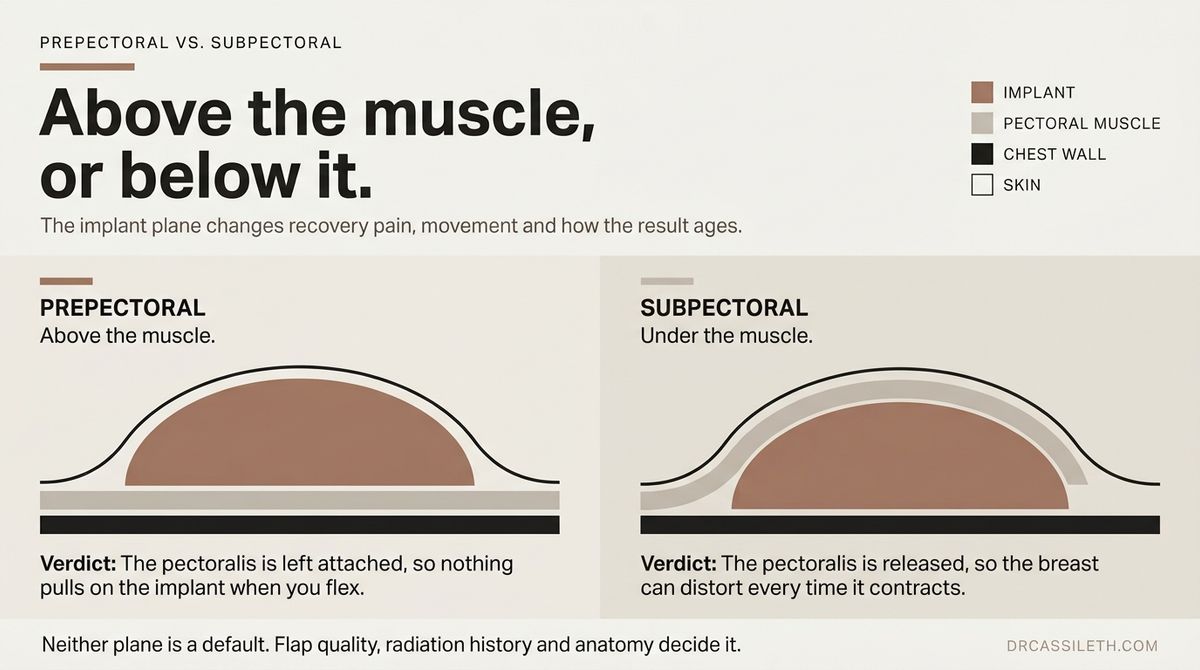
Prepectoral breast reconstruction places the implant in front of the pectoralis major, directly beneath the mastectomy skin. That is the same space your breast tissue filled before surgery, and that is the entire point.
Because no muscle covers the implant, support has to come from somewhere else. Most surgeons wrap it in acellular dermal matrix, then tuck it under the skin flap. Dr. Cassileth instead builds a full internal bra from that matrix, so your implant sits in a supported slope rather than sinking to the bottom of the pocket.
Meanwhile, your muscle is left untouched. You feel that in week one, and again with every push-up after. See her full reconstruction options.
What Is Subpectoral Breast Reconstruction?
Subpectoral breast reconstruction places the implant partly or fully beneath the pectoralis major. To build that pocket, your surgeon releases the lower border of the muscle from your chest wall, then covers the bottom of the implant with ADM.
For four decades, this was the standard, and for good reason. Mastectomy technique routinely left thin, poorly supplied skin back then, so the muscle worked as a second layer of insurance over the implant.
You pay for that coverage twice, though. Detaching the pectoralis hurts, sometimes for months.
And because the muscle now lies across your implant, it moves the breast whenever it contracts, lifting or flattening it when you push, lift, or reach overhead. Across 3,101 patients pooled in Annals of Surgical Oncology, that risk was dramatically lower without muscle over the implant.
Prepectoral vs. Subpectoral Breast Reconstruction: Key Differences
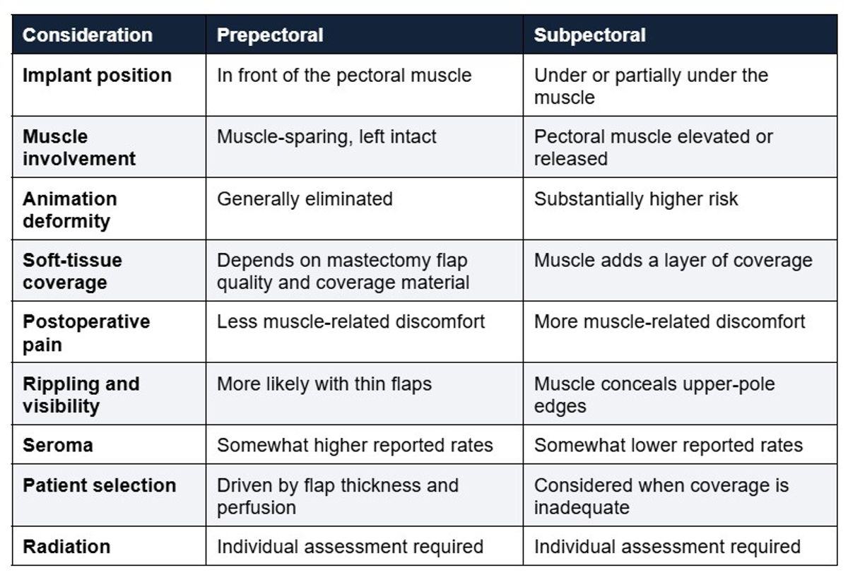
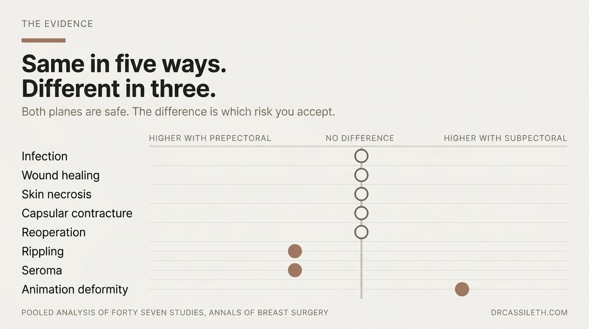
So how settled is that comparison? A 2025 meta-analysis in Annals of Breast Surgery pooled 47 studies covering 8,350 patients and found no meaningful difference between the two planes of implant-based breast reconstruction in infection, wound healing, necrosis, capsular contracture, or reoperation.
Only three outcomes differed. Prepectoral breast reconstruction showed more seroma and rippling, while subpectoral showed considerably more animation deformity.
That is good news for you. Because both approaches are safe, the decision comes down to which trade-off suits your body and your life, which is exactly the conversation your surgeon should be having with you.
Potential Benefits of Prepectoral Breast Reconstruction
Muscle preservation is the headline benefit of prepectoral breast reconstruction, and it is not a small one. Your pectoralis pushes, lifts, and stabilizes your shoulder, so leaving it attached means it keeps doing that job.
The evidence tracks with that. Reviewing 1,557 reconstructions, Frontiers in Oncology recorded fewer overall complications with prepectoral placement, 23.9% against 31.4%, plus less than half the rate of hematoma.
Animation deformity, though, is the clearest win. Across 227 prepectoral breasts, reviewers found zero cases, because a muscle that was never released cannot pull on your implant.
Capsular contracture came out comparable. And should it develop, capsular contracture treatment is one of the cases Dr. Cassileth's practice is built around, with a 95% success rate.
Potential Limitations and Risks of Prepectoral Reconstruction

Prepectoral is not a straight upgrade. It trades one set of problems for another, and you should know which set you are choosing.
Soft-Tissue Coverage and Implant Visibility
Nothing hides the implant in prepectoral breast reconstruction except your own skin and the matrix beneath it. That is why the thickness of your mastectomy flaps matters so much here, because thin flaps let the implant edge read through as visible ripples along the upper breast.
The Annals of Breast Surgery meta-analysis put that risk at roughly twice the subpectoral rate.
It can also be reduced up front. Dr. Cassileth's internal bra holds the implant against the chest wall in a supported slope, softening its contour and reducing how much fat grafting you need later.
Fat grafting handles the rest, thickening the layer over the implant, which is why it anchors most breast reconstruction revision work.
Surgical Complications
Both planes carry the same core risks, from infection to fluid collection to skin healing problems, and the plane you choose barely moves those numbers.
What does move them is size. Reviewing 499 consecutive prepectoral breast reconstructions in her own published series in Aesthetic Surgery Journal Open Forum, Dr. Cassileth found skin flap necrosis in 3.8% of cases overall. Implants above 500 cc, however, pushed that to 9.6%, against 2.3% below the threshold. Seroma and implant loss also climbed with size.
Notably, going larger than your original breast did not raise risk. Absolute volume did.
So the conversation worth having is not only about the plane. It is about how much implant your skin can safely carry.
Who May Be a Candidate for Prepectoral Reconstruction?
Candidacy for breast reconstruction after mastectomy comes down to your flaps. Reviewers place the working threshold near a centimeter of well-perfused tissue, and no chart predicts that in advance.
Several things narrow your odds:
- Active smoking, which constricts the vessels feeding your flap.
- High BMI, an independent predictor of skin necrosis.
- Poorly controlled diabetes or cardiovascular disease.
- Prior radiation, which stiffens skin.
- Thin flaps or visible ischemia during surgery.
Still, the biggest variable is not you. It is your mastectomy surgeon's technique, which is why Dr. Cassileth chooses hers carefully.
How Radiation Therapy Can Affect the Decision
Radiation is where the usual advice most often misses. The assumption is that irradiated skin needs muscle backing, so radiation sends you subpectoral by default.
The evidence complicates that. Pooling 14 studies of patients receiving postmastectomy radiotherapy, the Journal of Plastic, Reconstructive & Aesthetic Surgery found comparable complication rates and severity across both planes.
Dr. Cassileth goes further, favoring prepectoral breast reconstruction in previously radiated breasts, since it avoids the repeat surgeries that raise infection risk in fragile tissue.
Either way, raise your radiation plan with both your oncology team and your surgeon before the plane is chosen, not after.
The Role of Acellular Dermal Matrix and Other Coverage Materials
Acellular dermal matrix is donated human or animal dermis stripped of its cells, leaving a collagen scaffold your own tissue grows into. In prepectoral breast reconstruction, it takes over the job your pectoralis used to do, supporting the implant, defining the pocket, and adding a layer between the device and your skin.
Synthetic meshes do similar work, though what separates them matters to you:
- Acellular dermal matrix, which your own tissue gradually integrates into.
- Poly-4-hydroxybutyrate mesh, absorbable, dissolving as your tissue takes over the support.
- Titanium-coated polypropylene, permanent, staying in the body for good.
That said, no material is mandatory, and evidence has not shown a universal complication benefit from ADM. What separates results is technique. Wrapping an implant and tucking it under the skin is not the same operation as building a supported internal bra from the same product.
Prepectoral vs. Subpectoral Breast Reconstruction: Which Is Better for You?
Patient or Surgical Factor
What to Discuss
Mastectomy-flap quality
Thickness, blood supply, healing potential
Radiation
Previous exposure or anticipated treatment
Smoking
Impact on healing and complication risk
Body composition
Available soft-tissue coverage
Breast size and shape
Desired result and implant coverage
Chest muscle activity
How much avoiding animation deformity matters to you
Implant visibility concerns
Amount and quality of soft-tissue coverage
Prior augmentation
Existing pocket often favors subpectoral placement
Cancer treatment plan
Timing and type of reconstruction
Notice what the table cannot do: rank these factors for you. Your surgeon weighs them together, and often the final call comes in the operating room. Across 576 reconstructions in the Journal of Clinical Medicine, surgeons chose the plane after the mastectomy, once the defect's shape was visible.
Patients there also reported higher satisfaction with prepectoral placement.
One factor gets overlooked, though. If you already have implants, your muscle is usually partly detached, which typically pushes you toward subpectoral breast reconstruction.
What to Expect During the Reconstruction Process
Two decisions shape your reconstruction, and they are easy to confuse. One is plane, meaning where the implant sits relative to your chest muscle. The other is timing, meaning when the reconstruction happens and how many operations it takes.
They combine independently, so settling one does not settle the other.
Immediate vs. Delayed Reconstruction
Immediate reconstruction happens during the mastectomy itself, in one operation. Because your breast skin is never left empty, it keeps its shape instead of tightening and shrinking over the chest wall.
Dr. Cassileth performs most of her reconstructions this way, coordinating directly with the breast surgeon so both procedures run as one.
Delayed reconstruction comes later, often after chemotherapy or radiation finishes. Surgeons choose it when the treatment sequence requires it, or when irradiated tissue needs months to recover before it can safely hold an implant.
That distinction matters less than you might expect, though, because your oncology timeline usually decides it for you.
Tissue Expander vs. Direct-to-Implant Reconstruction
Staged reconstruction starts with a tissue expander, a temporary device your surgeon inflates over roughly three months to stretch the skin, then swaps for a permanent implant in a second surgery. Direct-to-implant skips that entirely, placing the final implant during your mastectomy.
So the real question is whether your skin needs stretching at all. Prepectoral breast reconstruction sits the implant where breast tissue already was, in a space that exists the moment the mastectomy ends.
That is the pairing Dr. Cassileth built direct-to-implant around, which is why expanders rarely enter her cases.
Questions to Ask Your Breast Reconstruction Surgeon
Bring these to your consultation, because the answers reveal as much about your surgeon as about the surgery:
- Given my flap quality, am I a candidate for prepectoral breast reconstruction?
- Who is performing my mastectomy, and what is that surgeon's flap necrosis rate?
- How does my radiation history change the plane you recommend?
- Expander or direct-to-implant, and why for me specifically?
- How do you correct rippling if it appears?
- How many of each do you perform yearly?
Push for answers built on your anatomy. A surgeon quoting only published averages has not examined you closely enough.
So Which Plane Fits Your Case?

Only an examination answers that. Prepectoral breast reconstruction spares your chest muscle, eliminates animation deformity, and eases recovery, but your flap quality, radiation history, and goals decide whether it suits you.
That judgment takes experience. Dr. Cassileth developed direct-to-implant reconstruction and has refined it over two decades, performing more than 500 reconstructions a year.
If you are weighing implant-based breast reconstruction, book a consultation in Beverly Hills and get an answer built around your anatomy.
FAQs
What is the difference between prepectoral vs subpectoral breast reconstruction?
Placement. Prepectoral breast reconstruction sits the implant in front of your chest muscle, where breast tissue used to be. Subpectoral breast reconstruction places it partly beneath the muscle, which the surgeon must release.
That single difference drives recovery pain, animation deformity, and how much coverage sits over your implant.
Can prepectoral reconstruction be performed after radiation therapy?
Often yes. Recent pooled evidence found comparable complication rates and severity between both planes in patients receiving postmastectomy radiotherapy.
Radiated skin still requires individual assessment of thickness and perfusion, so bring your full radiation history, past or planned, to your reconstructive consultation before any decision about plane is made.
Does prepectoral reconstruction eliminate animation deformity?
Essentially yes. Animation deformity happens when a pectoral muscle covering your implant contracts and distorts the breast. Because prepectoral placement leaves that muscle untouched, rates drop dramatically.
If you already have animation deformity from implant-based breast reconstruction, conversion to a prepectoral pocket is sometimes possible.
How long does prepectoral breast reconstruction take to heal?
Most patients spend one night in hospital and return to desk work within roughly two weeks. Drains usually come out in seven to ten days.
Full activity resumes near six weeks, though your final shape keeps settling for months. Sparing the muscle generally makes those early weeks more comfortable.
Can breast reconstruction after mastectomy be revised later?
Yes, and revision is normal, not a sign of failure. Fat grafting softens rippling and contour irregularities, implants can be exchanged for a different size or profile, and capsule work corrects contracture or malposition.
What suits you depends on your tissue quality and the original surgical technique.
More from the journal


